File:MYB FISH break-apart probe.png
Jump to navigation
Jump to search
No higher resolution available.
MYB_FISH_break-apart_probe.png (497 × 466 pixels, file size: 162 KB, MIME type: image/png)
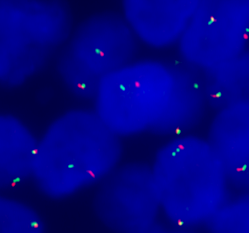
FISH with a break-apart probe for 5' and 3' MYB gene region shows separate red and green probe signals, consistent with MYB rearrangement. The separation in this case was small rather than a wide split between 5' and 3' MYB gene regions, suggesting that the mechanism of rearrangement was an inversion (intrachromosomal rearrangement).
File history
Click on a date/time to view the file as it appeared at that time.
| Date/Time | Thumbnail | Dimensions | User | Comment | |
|---|---|---|---|---|---|
| current | 17:22, 9 November 2023 | | 497 × 466 (162 KB) | Kgeiersbach (talk | contribs) |
You cannot overwrite this file.
File usage
The following page uses this file: